
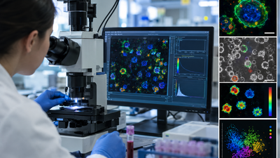
A routine blood sample contains a surprisingly detailed record of what the immune system is doing. Scientists have now demonstrated a way to read some of that information without adding fluorescent dyes or other chemical labels. The approach examines the natural light emitted by molecules inside immune cells, allowing researchers to assess their metabolism at the single-cell level. In experiments, the technique helped distinguish activated immune cells from resting ones with nearly 94% accuracy just two hours after stimulation. According to the study published in Biophotonics Discovery, researchers used autofluorescence lifetime imaging to examine peripheral blood mononuclear cells (PBMCs) and assess their metabolic characteristics at the single-cell level. The technique relies on naturally occurring cellular fluorescence, allowing immune cells to be studied without the need for added fluorescent dyes.
Reading cells without dyes
Scientists commonly use fluorescent antibodies and other labels to identify immune cells by looking for particular surface markers. These methods are well established, but they require additional reagents and preparation steps. They can also provide relatively limited information about what individual cells are actually doing. The new approach takes a different route. Instead of attaching a fluorescent marker to a cell, researchers measure its autofluorescence; light naturally produced by molecules inside cells.The technique is based on optical metabolic imaging (OMI), which uses two-photon microscopy to excite naturally fluorescent molecules involved in cellular metabolism. Researchers then measure the fluorescence lifetime, or how long the molecules remain in an excited state before returning to their normal state. Those measurements can provide clues about a cell’s metabolic condition. Because the method relies on signals already present inside the cells, researchers can examine them without introducing an external dye.
Thousands of cells analysed
The researchers tested the method using PBMC samples obtained from three healthy donors. PBMCs include several important immune-cell populations, including T cells, B cells, monocytes and natural killer (NK) cells. The team examined thousands of individual cells under both resting and activated conditions. Machine-learning algorithms were then used to determine whether the metabolic information captured through imaging could distinguish immune-cell types and reveal whether cells had become activated.One of the most notable findings involved the difference between resting and activated cells. The imaging approach distinguished activated PBMCs from resting cells with nearly 94% accuracy only two hours after stimulation. That is important because immune activation can involve rapid changes in cellular metabolism. Rather than simply determining which cells are present, the method can provide information about their functional state.
Different cells leave different signals
The researchers also found that individual immune-cell populations had distinct metabolic patterns. Monocytes, which are part of the innate immune system and can respond quickly to threats, were identified with 96% accuracy in resting samples and 88% accuracy after activation. NK cells were identified with about 74% accuracy in both resting and activated conditions.The results were less clearly separated for some adaptive immune cells. T cells and B cells showed more similar metabolic profiles under the experimental conditions. This highlights an important feature of the technique: cellular metabolism can act as an additional source of information alongside conventional markers. Two cells that appear similar based on their identity may not necessarily have the same metabolic state.
Single-cell analysis reveals hidden variation
Looking at individual cells also exposed differences that could be missed when researchers analyse an entire population as a single sample. Within the same immune-cell population, some cells showed relatively high metabolic activity while others remained comparatively quiet. A bulk measurement would average these signals together, potentially hiding that variation.Single-cell imaging, by contrast, allows researchers to see the differences from one cell to another. That could be particularly useful when studying immune responses, where only a fraction of cells may respond strongly to a stimulus. The ability to make these measurements without relying on fluorescent labels for the imaging itself could also have applications beyond basic research. PBMCs are already used in research and clinical workflows and can serve as starting material for immune-cell therapies. A non-destructive way of assessing cellular condition could eventually help researchers evaluate the quality or fitness of cells before further processing.
Potential for immune research
The researchers caution that the technique is not yet a replacement for established cell-identification methods. Its accuracy varies between immune-cell populations, and conventional fluorescent labelling remains more effective for identifying many specific cell types. Its strength lies elsewhere: the method provides functional metabolic information at single-cell resolution without requiring external fluorescent labels. That could make it a useful complement to existing approaches for studying diseases in which immune activity and cellular metabolism change, including cancer and immune disorders. It may also be valuable for research into cell-based therapies, where understanding the condition of individual cells could be important.The study demonstrates how optical imaging can move beyond simply showing what cells look like. By analysing the natural fluorescence produced by cellular metabolism, researchers can begin to uncover how individual immune cells behave, and detect metabolic changes associated with activation surprisingly early after stimulation.